

When it comes to spinal health, few physicians understand both sides of the story like Dr. Todd Lanman. As a renowned spinal neurosurgeon practicing in Beverly Hills and affiliated with Cedar-Sinai and UCLA medical centers, Dr. Lanman brings a unique perspective to treating disc conditions—he’s not only a skilled surgeon who has overseen numerous clinical trials for artificial disc replacement, but also a patient who has personally experienced the journey back to spine health having undergone 11 spinal surgeries himself. This firsthand experience makes him especially attuned to the challenges of conditions like disc extrusion.
Disc extrusion is a specific type of herniated disc where the inner gel-like material (nucleus pulposus) breaks through the outer fibrous ring (annulus fibrosus) though it remains connected to the disc. Unlike the milder disc protrusion where the nucleus pushes against but doesn’t break through the annulus, an extrusion represents a more severe condition. Extruded material can compress nearby nerves causing significant pain, numbness, and weakness.
Early diagnosis of extrusion disc herniation is important – delays can lead to chronic pain, permanent nerve damage, muscle weakness, and even loss of mobility. Whether through conservative management like physical therapy and pain medication or surgical interventions such as microdiscectomy or artificial disc replacement, addressing disc extrusion promptly can prevent these serious long-term complications and help restore quality of life.
Understanding Extrusion Disc Herniation
What Is an Extrusion Disc Herniation?
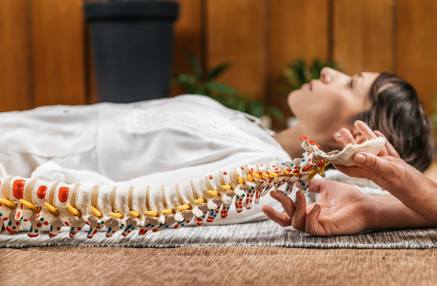
A disc herniation occurs when the soft, gel-like center of a spinal disc (nucleus pulposus) pushes through a tear in the tough, fibrous outer layer (annulus fibrosus). In an extrusion disc herniation specifically, the nucleus material not only breaks completely through the outer annulus wall, but the depth of the herniated material is larger than its width. This makes it distinctly different from a disc protrusion (or bulging disc), where the nucleus pushes against and stretches the annulus but doesn’t break through it completely. Think of a protrusion like a balloon being squeezed on one side, creating a bulge, while an extrusion is more like toothpaste being squeezed through a tear in the tube.
What makes disc extrusion unique is that while the nucleus material escapes through tears or fissures in the annulus fibrosus, it remains connected to the main disc. This connection to the original disc distinguishes it from the more severe disc sequestration, where the nucleus material completely separates from the disc and floats freely in the spinal canal. The extrusion process often begins as a protrusion that progressively worsens as pressure continues to push against a weakened annulus wall until it finally gives way. Once the annulus is torn and the nucleus material extrudes, the disc becomes vulnerable to further damage since these annular tears cannot heal spontaneously due to the disc’s poor blood supply.
Causes of Extrusion Disc Herniation
Disc degeneration is a common process that begins early in life, often starting in the second decade. As we age, the amount of water in our vertebral discs decreases, making them less flexible and more prone to tearing. This age-related wear and tear, known as disc degeneration, is the most common cause of disc herniation, including extrusion.
Occupational and Physical Factors
Certain occupations and physical activities significantly increase the risk of developing disc extrusion. Occupational tasks including physical overload, handling heavy loads, and self-reported hard work are all associated with increased risk of lumbar disc herniation. Workers exposed to biomechanical overload were found to have a nearly four times higher risk of lumbar disc herniation compared to those not exposed.
Traumatic Injuries
While less common than degenerative causes, traumatic events can directly cause disc extrusion. Traumatic lumbar disc herniation can result from accidents, falls, or sudden impacts. In one documented case, a patient developed a stenosing traumatic lumbar disc herniation with a large sequestration after being thrown from a horse. Traumatic disc herniations can cause symptoms similar to degenerative disc herniations, including low back pain, sciatica, paresthesia, and muscle weakness in the legs.
Genetic Predisposition
Recent research has significantly shifted our understanding of disc herniation causes, highlighting the substantial role of genetic factors. Twin studies have revealed that genetic factors account for up to 75% of individual susceptibility to intervertebral disc degeneration, with environmental factors having a more modest impact than previously thought. The Twin Spine Study, which followed twins from Canada, Finland, and the United States, found that identical twins with different jobs, physical activities, and interests demonstrated similar levels of disc degeneration, suggesting genetic factors have a much larger influence than expected.
Common Symptoms of Extrusion Disc Herniation
Extrusion disc herniation typically produces a range of symptoms that can significantly impact daily life. Unlike a bulging disc, where symptoms may be mild or absent, an extruded disc often causes more pronounced symptoms due to the nucleus material breaking through the outer annulus and potentially compressing nearby nerves.
Localized Pain
Pain at the site of the herniated disc is common, particularly in the lower back for lumbar herniations or neck for cervical herniations. This pain occurs due to the displaced disc fragment pressing on spinal nerves. The pain is usually continuous or at least continuous in specific body positions, unlike the pulsating or intermittent pain caused by muscle spasms.
Radiating Pain
One of the most distinctive symptoms is pain that radiates along nerve pathways:
- Sciatica: When a lumbar disc extrudes, it often causes sharp, shooting pain that radiates from the buttocks down the leg due to pressure on the sciatic nerve. This pain is frequently described as sharp and electric shock-like, and may worsen with standing, walking, or sitting.
- Cervical Radiculopathy: With cervical disc extrusion, pain can begin in one area, such as the shoulder, and travel along the arm to the fingers. Patients with cervical radiculopathy may experience neck pain radiating to the upper and lower limbs.
Numbness and Tingling
Sensory changes are common when an extruded disc compresses nerve roots:
- Paresthesia (abnormal sensations like “pins and needles”)
- Numbness or tingling in the limbs
- Hypoesthesia (reduced sensation) in the area served by the affected nerve
These sensory symptoms typically follow the distribution pattern of the compressed nerve. For example, a C5/C6/C7 disc extrusion might cause reduced sensation in those specific dermatomes.
Weakness or Loss of Function
Muscle weakness occurs when the extruded disc material puts pressure on the spinal cord or nerve roots, making it harder for nerves to carry signals to and from the brain:
- Reduced grip strength or weakness in the extremities for cervical herniations
- Weakness in the leg or foot for lumbar herniations
- Difficulty walking, especially with thoracic disc herniations
- In severe cases, progressive weakness in lower extremities
In rare but serious cases, disc extrusion can lead to bowel and bladder dysfunction if it causes significant compression of the spinal nerve roots or cauda equina. This represents a medical emergency that requires immediate attention.
Importantly, symptoms vary widely between individuals. Some people with disc extrusion might experience all these symptoms, while others might have only one or two predominant symptoms. In rare cases, a person might have a disc extrusion with no noticeable symptoms if the extruded material does not press on soft tissues or nerves.
Diagnosis and Medical Evaluation
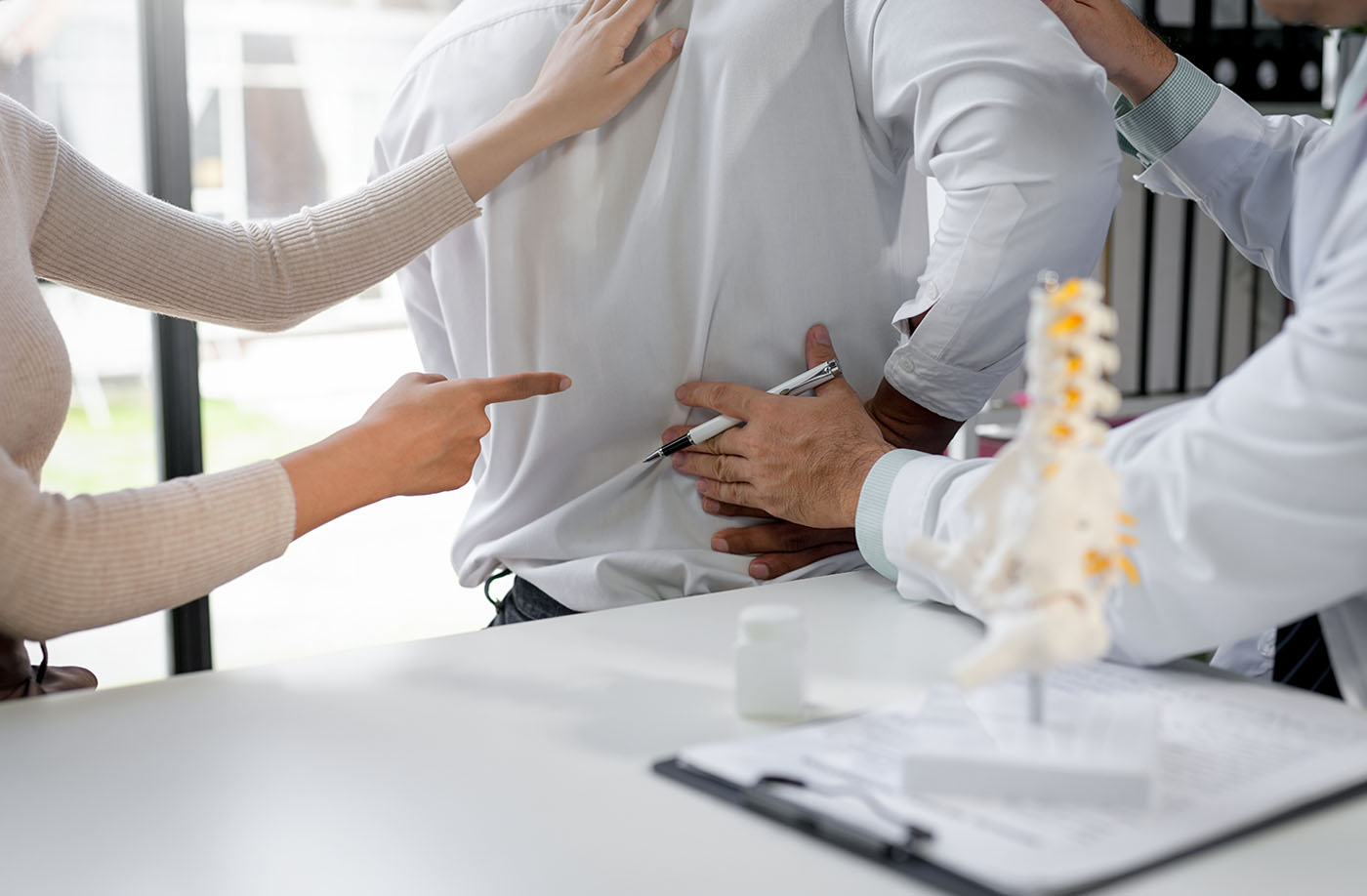
How Is an Extruded Disc Diagnosed?
Diagnosing an extruded disc involves a comprehensive approach that combines physical examination, imaging tests, and neurological assessment. This multi-faceted diagnostic process helps healthcare providers accurately identify the condition and determine its severity.
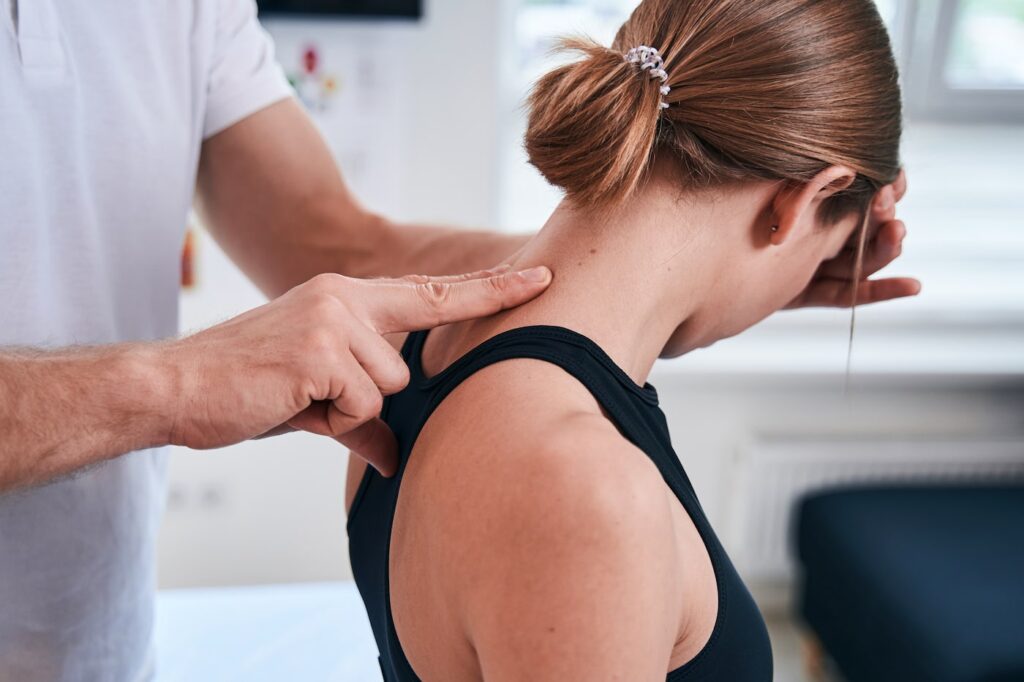
Physical Examination
The physical examination is a crucial first step in diagnosing an extruded disc:
- Doctors assess mobility limitations, balance problems, and pain levels.
- The straight leg raise test is particularly useful for diagnosing lumbar disc herniation. If raising the leg induces or worsens pain, it suggests sciatic nerve root compression.
- Muscle strength, reflexes, and any visible muscle twitches or spasms are evaluated.
Imaging Tests
Imaging tests provide detailed visual evidence of disc herniation:
- Magnetic Resonance Imaging (MRI) is the gold standard for diagnosing extruded discs. It offers the most accurate assessment of herniation location and affected neural tissue.
- Computed Tomography (CT) scans, especially when combined with myelography, can be used when MRI is not possible.
- X-rays, while not able to show disc material directly, can help rule out other conditions like vertebral fractures or spondylolisthesis.
Neurological Assessment
A thorough neurological evaluation helps determine the extent of nerve involvement:
- Doctors test for unresponsive nerve reflexes in various body parts using tools like a reflex hammer.
- Sensory tests assess skin sensitivity to stimuli like temperature changes.
- Specific muscle groups are evaluated for strengths and weaknesses.
In some cases, additional tests may be performed:
- Electromyography (EMG) measures electrical impulses along nerves and muscles, helping determine if pain or weakness originates from nerves or muscles.
- Nerve conduction studies can be used to assess the severity and progression of nerve damage.
Non-Surgical Treatment Options for Extrusion Disc Herniation
Conservative Pain Management
Conservative pain management for extrusion disc herniation typically involves a combination of physical therapy, medication, and epidural steroid injections. These non-surgical approaches aim to alleviate pain, reduce inflammation, and improve function without resorting to invasive procedures.
- Physical Therapy: Physical therapy plays a crucial role in managing herniated disc pain and promoting healing. Strengthening exercises focus on building core and back muscles to support the spine. Posture correction techniques help reduce pressure on the affected disc. Specific exercises and stretches are tailored to each patient’s condition. Physical therapists create personalized treatment plans to address individual needs and symptoms.
- Medication: Various medications can help manage pain and inflammation associated with disc extrusion. Anti-inflammatory drugs (NSAIDs) like ibuprofen or naproxen reduce inflammation and pain. Muscle relaxants may be prescribed to alleviate muscle spasms often accompanying disc herniation. Pain relievers, ranging from over-the-counter acetaminophen to prescription opioids, can be used for pain management. In some cases, oral steroids may be prescribed for short-term use to reduce swelling.
- Epidural Steroid Injections: Epidural steroid injections are a targeted approach to pain relief. Injections deliver corticosteroids directly into the epidural space around the spinal cord. These injections reduce inflammation and pressure on irritated nerve roots. Injections can provide temporary pain relief, allowing time for the nerve to heal. They are typically administered no more than three times per year. Steroid injections can be particularly effective for radicular pain caused by nerve root compression.
Lifestyle Modifications for Disc Health
By making thoughtful changes to your daily habits and environment, you can significantly reduce strain on your spine and promote healing.
- Ergonomic changes for work and daily activities: Ergonomics refers to designing and arranging workspaces, tools, and equipment to promote efficiency, comfort, and safety. Proper ergonomics is crucial for maintaining good health and preventing injuries like herniated discs. To incorporate ergonomics into your workspace, adjust your chair and desk height, use an ergonomic keyboard and mouse, ensure your monitor is at eye level, and position frequently used items within easy reach. For proper posture while sitting, ensure your feet are flat on the floor, your back is supported by the chair, and your shoulders are relaxed. When standing, distribute your body weight evenly on both feet and keep your shoulders back. Taking regular breaks every 30-45 minutes to stand, stretch, and walk around can help prevent prolonged sitting, which increases pressure on spinal discs.
- Weight management and proper exercise routines: Excess weight places additional strain on your spine and can contribute to disc herniation. Weight loss achieved through diet and exercise can significantly reduce the burden on your body. Losing weight decreases the load on the spine, relieving pressure on the spinal discs and joints, which can help alleviate back pain. Additionally, weight loss can decrease the rate of disc degeneration, reducing the risk of herniated discs and spinal stenosis.
Surgical Treatments for Extrusion Disc Herniation
When it comes to treating extrusion disc herniation, surgery becomes a consideration when conservative treatments fail to provide relief or when certain serious conditions develop. Understanding when surgery is appropriate can help patients make informed decisions about their care.
When Is Surgery Needed?
- Persistent pain despite conservative treatments: Surgery may be recommended when a patient experiences continued pain that doesn’t respond to non-surgical treatments after about six weeks. Studies show that for symptoms resistant to initial conservative treatments, discectomy to remove the offending herniated disc material may provide faster and greater pain relief compared to continued non-surgical care. Many patients report marked improvements to their pain symptoms in as little as one week following surgery.
- Progressive nerve damage or loss of function: Surgery becomes necessary when there is evidence of worsening neurological symptoms or deficits, such as increasing weakness, numbness, or difficulty walking. Early surgical intervention in these cases can help prevent permanent nerve damage and may facilitate recovery of function. This is particularly important when diagnostic imaging confirms disc extrusion that corresponds with clinical findings.
- Severe spinal instability or compression: Absolute surgical indications include deteriorating neurological deficits with myelopathy or cauda equina syndrome. Cauda equina syndrome is characterized by lower back pain, paralysis of leg muscles, loss of bowel and bladder control, and numbness around the groin. These conditions are medical emergencies requiring immediate surgical intervention.
Surgical Options for Disc Herniation
Common surgical procedures for disc extrusion include:
- Microdiscectomy: This minimally invasive procedure involves removing the herniated disc material through a small incision. Using a microscope or endoscope for enhanced visualization, the surgeon can precisely target and remove the extruded disc fragment while minimizing damage to surrounding tissues. Microdiscectomy typically results in faster recovery times and less postoperative pain compared to traditional open discectomy.
- Laminectomy: In this procedure, surgeons remove part of the vertebral bone called the lamina to decompress nerves. By creating more space within the spinal canal, a laminectomy relieves pressure on the spinal cord or nerve roots. This can be particularly effective for treating spinal stenosis that may accompany disc extrusion. While more invasive than microdiscectomy, laminectomy may be necessary when multiple levels of the spine are affected or when there’s significant canal narrowing.
- Artificial Disc Replacement (ADR): ADR is a motion-preserving alternative to spinal fusion for certain patients. In this procedure, the damaged disc is removed and replaced with an artificial disc device. This approach aims to maintain normal spine movement and potentially reduce the risk of adjacent segment degeneration often associated with fusion surgery. ADR is particularly beneficial for younger, active patients with single-level disc disease and no significant facet joint arthritis.
- Spinal Fusion: While used more selectively due to its impact on spinal mobility, fusion remains an important option for cases of severe disc damage or instability. This procedure involves joining two or more vertebrae together using bone grafts and sometimes metal implants. Fusion can provide stability and pain relief but at the cost of some spinal flexibility. It’s typically reserved for cases where disc degeneration is advanced, there’s significant instability, or when other surgical options are not appropriate.
Recovery and Long-Term Management
Recovery time varies but typically ranges from 6 to 12 weeks, depending on the type of treatment. Most people can return to work after 2-4 weeks, though those with physically demanding jobs may need 12 weeks or more.
Post-Treatment Rehabilitation
- Gradual return to activity and physical therapy: Most people begin to see improvement within a few weeks of starting physical therapy. Full recovery may take several months. Light activity should resume soon after initial treatment to help discs heal and absorb nutrients.
- Pain management strategies: May include over-the-counter pain relievers, anti-inflammatory drugs, or prescription medications. Physical therapy, alternative therapies like acupuncture, and mindfulness techniques can also help manage pain long-term.
Preventing Future Disc Herniation
- Core strengthening exercises: Focus on exercises that engage core muscles to improve spine stability such as planks, bird dogs, and glute bridges.
- Proper body mechanics and posture: Maintain good posture, use proper lifting techniques, and set up an ergonomic workspace to reduce stress on the spine.
Additional preventive measures include maintaining a healthy weight, taking regular breaks from prolonged sitting, wearing supportive shoes, and quitting smoking.
Why Choose Dr. Todd Lanman for Extrusion Disc Herniation Treatment?
Dr. Lanman is recognized globally as a leading authority in minimally invasive spine surgery. Rather than creating large incisions, he uses advanced techniques requiring only small incisions—typically about an inch long. Through these small openings, he can insert specialized instruments and cameras (endoscopes) to access and treat the affected area of your spine. This approach reduces muscle damage, decreases recovery time, and leads to better outcomes for patients.
He takes a holistic approach to spinal care through his proprietary 4D Health Process. He examines all aspects of a patient’s health—including age, lifestyle, nutrition, hormone status, physical exercise routine, and spine and joint health—to create comprehensive, personalized treatment plans. This multidimensional approach ensures that treatment addresses not just the immediate disc problem but your overall well-being and quality of life.
Dr. Lanman uses cutting-edge technology for both diagnosis and treatment, including intraoperative fluoroscopy and three-dimensional CAT scans. These technologies allow for more accurate placement of artificial discs and other implants.
Don’t let extrusion disc herniation limit your life any longer. Schedule a consultation with Dr. Todd Lanman today at his Beverly Hills location. Take the first step toward a pain-free, active lifestyle by calling his office or making an appointment online.
FAQs: Extrusion Disc Herniation
How serious is an extruded disc?
An extruded disc is a more severe form of disc herniation, where the inner gel-like material pushes through the outer layer and may press on nearby nerves. It can cause significant pain, numbness, or weakness, especially if nerve roots are compressed. In some cases, it may require medical or surgical treatment.
Can an extruded disc heal without surgery?
Yes, many extruded discs improve with conservative treatment such as physical therapy, anti-inflammatory medications, and activity modification. The body can gradually reabsorb part of the disc material, reducing pressure on nerves. However, if symptoms persist or worsen, surgery may be necessary.
How long does it take to recover from an extruded disc?
Recovery time varies but often takes several weeks to a few months with non-surgical treatment. Most people improve within 6 to 12 weeks with rest, physical therapy, and medications. Surgical recovery may be quicker for some, but depends on the procedure and individual health factors.
What’s the difference between an extruded and a bulging disc?
A bulging disc occurs when the disc’s outer layer extends outward but remains intact. An extruded disc is more severe—part of the inner disc material breaks through the outer layer and may press on nerves. Extrusions are more likely to cause significant symptoms and may require more intensive treatment.
When should I see a spine specialist for a herniated disc?
See a spine specialist if you have persistent back or neck pain, numbness, weakness, or pain radiating into the arms or legs. Immediate evaluation is needed if you experience loss of bladder or bowel control, severe weakness, or difficulty walking, as these may indicate a serious condition.