

Spinal bone spurs, or osteophytes, often sound more alarming than they are. These bony projections form as the spine adapts to years of movement, stress, and natural wear. In many people, they cause no symptoms at all—but when they encroach on nearby nerves or the spinal cord, they can trigger sharp pain, stiffness, and even weakness that limits daily life. Understanding why bone spurs form and when they become a problem is the first step toward relief. With advanced motion-preserving treatments now available, patients no longer have to accept chronic discomfort or reduced mobility as part of aging.
This guide explains what spinal bone spurs are, why they form, the symptoms to watch for, and when treatment is necessary. You’ll also learn how advanced, motion-preserving techniques at ADR Spine can help restore mobility and long-term spinal health.
Understanding Spinal Bone Spurs
What Are Bone Spurs?
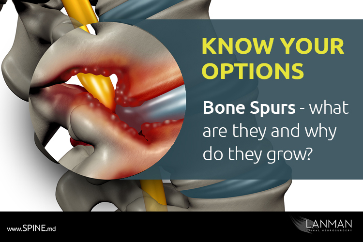
Bone spurs, medically known as osteophytes, are small bony projections that develop along the edges of joints or vertebrae. They form as the body’s attempt to stabilize areas affected by inflammation, mechanical stress, or age-related degeneration. Over time, as the cushioning discs and cartilage between spinal joints wear down, the body responds by producing extra bone to reinforce those weakened areas. While this process is meant to protect the spine, the resulting bone spurs can narrow the spaces where nerves travel, leading to pain or restricted movement when they press on nearby structures.
Where Bone Spurs Occur in the Spine
Bone spurs can develop anywhere along the spine, but they most often appear in the cervical (neck), thoracic (mid-back), and lumbar (lower back) regions. In the neck, they may press on nerves that extend into the shoulders and arms, causing pain or tingling. In the lower back, they can irritate nerves that travel to the hips or legs, sometimes mimicking sciatica. Even in the mid-back, where the spine is more stable, bone spurs can restrict movement or cause localized discomfort. Their impact depends on both their size and location; some bone spurs remain harmless, while others interfere with the delicate balance of spinal mobility and nerve function.
Why the Spine Develops Bone Spurs
Bone spurs form as part of the spine’s natural repair response to instability or degeneration. When spinal discs thin or joints lose cartilage, the body senses excess motion and friction between vertebrae. To protect these weakened areas, it deposits new bone along the edges of the vertebrae, creating osteophytes. Initially, this extra bone may stabilize the joint, but over time it can encroach on nerve pathways or the spinal canal. The result is often the opposite of what the body intended—nerve compression that causes pain, stiffness, or radiating symptoms into the arms or legs.
Causes of Spinal Bone Spurs
Degenerative Disc Disease (DDD)
Degenerative disc disease occurs when the soft, cushioning discs between vertebrae lose height and hydration over time. As these discs wear down, the space between vertebrae narrows, creating abnormal motion and instability in the joints of the spine. In response, the body tries to reinforce the area by forming new bone along the vertebral edges. These bony overgrowths help stiffen the joint but can also intrude into nearby nerve spaces. The result is a gradual shift from a protective process to one that contributes to pain, stiffness, and reduced flexibility.
Osteoarthritis and Aging
As the spine ages, the cartilage that cushions its joints naturally thins and becomes less resilient. This gradual breakdown, a hallmark of osteoarthritis, exposes bone surfaces to increased friction and inflammation. In an effort to stabilize the affected joints, the body produces new bone along the vertebral edges. These bone spurs can help limit excessive motion but often come at a cost—narrowing the spaces around nerves and the spinal cord. Over time, what begins as a protective adaptation can contribute to chronic stiffness, pain, and nerve-related symptoms.
Poor Posture and Repetitive Stress
Years of poor posture or repetitive strain place continuous stress on the spine’s joints and supporting structures. Slouching, prolonged sitting, or repetitive lifting can cause uneven pressure on the vertebrae, accelerating wear in specific areas. The body responds to this chronic stress by forming extra bone to reinforce the overworked joints. While these bone spurs may initially offer support, they can eventually encroach on nerve pathways and contribute to pain, stiffness, or limited motion. Over time, even minor daily habits—such as leaning forward at a computer or carrying weight unevenly—can play a role in bone spur development.
Spinal Trauma or Injury
Spinal trauma, such as a fracture, dislocation, or whiplash injury, can disrupt the normal alignment and mechanics of the spine. Even after healing, the affected joints often bear abnormal stress that accelerates wear and degeneration. In response, the body may produce new bone in an attempt to stabilize the damaged area. Over time, these post-injury bone spurs can form near the site of trauma, where they sometimes irritate surrounding nerves or restrict movement. What begins as the body’s effort to restore stability can evolve into a source of persistent pain or stiffness years after the original injury.
Genetic and Lifestyle Factors
Genetic predisposition plays a meaningful role in how readily the body forms bone spurs. Individuals with a family history of osteoarthritis or spinal degeneration tend to develop osteophytes earlier or more extensively. Lifestyle factors can further heighten this risk. Excess body weight places added pressure on spinal joints, while physical inactivity weakens the muscles that support proper alignment. Together, these influences create mechanical stress that promotes cartilage breakdown and abnormal bone formation. Although genetics cannot be changed, maintaining a healthy weight, staying active, and using proper posture can help slow or prevent the progression of spinal bone spurs.
Symptoms of Spinal Bone Spurs
Localized Pain and Stiffness
Localized pain and stiffness are among the earliest signs of spinal bone spurs. Many patients describe a dull ache or tightness in the neck or lower back that worsens after periods of rest, such as first thing in the morning. This discomfort often eases somewhat with gentle movement, as motion temporarily relieves joint pressure. Over time, however, chronic inflammation and mechanical irritation from bone spurs can make the spine feel less flexible, turning routine movements—like bending, twisting, or standing up straight—into sources of pain.
Nerve Compression Symptoms
When bone spurs press on nearby spinal nerves, symptoms can extend far beyond the site of the spur. Patients may experience sharp or burning pain that radiates into the arms or legs, depending on where the compression occurs. Numbness, tingling, or a pins-and-needles sensation often accompany the pain, signaling irritation of sensory nerve fibers. In more advanced cases, muscle weakness or coordination problems can develop as the compressed nerves struggle to transmit signals properly. These nerve-related symptoms not only cause discomfort but can also interfere with fine motor control, balance, and overall mobility.
Reduced Range of Motion
Bone spurs can restrict normal joint movement by stiffening the spine and narrowing the spaces between vertebrae. Many patients notice difficulty turning their head, bending forward, or standing fully upright without discomfort. This loss of flexibility often develops gradually as the spine attempts to stabilize itself through new bone growth. Over time, the combination of joint stiffness and muscle tightness can make even simple motions like checking a blind spot while driving or reaching overhead feel strained or limited.
Spinal Cord Compression Signs (Advanced Cases)
In advanced cases, bone spurs can narrow the spinal canal itself, placing direct pressure on the spinal cord. This condition, called myelopathy, produces more serious neurological effects than routine nerve compression. Patients may develop unsteady balance, hand clumsiness, or difficulty performing fine motor tasks such as buttoning clothing or writing. In severe cases, bladder or bowel control may be lost. If you experience a loss of bladder or bowel control, seek medical attention immediately.
Diagnosing Spinal Bone Spurs
Physical and Neurological Exam
Diagnosis begins with a thorough physical and neurological examination. The physician evaluates posture, spinal alignment, and range of motion to identify areas of stiffness or abnormal curvature. Reflexes, muscle strength, and sensory responses are tested to detect nerve irritation or weakness caused by compression. Subtle findings such as decreased grip strength, delayed reflexes, or uneven muscle tone can point to specific spinal levels affected by bone spurs. Together, these clinical observations guide the decision to pursue imaging and help differentiate mechanical back pain from neurologic compromise.
Imaging Studies
Imaging studies provide the most definitive way to confirm the presence and severity of spinal bone spurs. X-rays can reveal the bony overgrowths themselves and show how they alter the shape or spacing of the vertebrae. Computed tomography (CT) offers more detailed visualization of bone anatomy, helping pinpoint spurs that encroach on nerve pathways. Magnetic resonance imaging (MRI) adds crucial information about the surrounding soft tissues, including discs, ligaments, and the spinal cord. Together, these tools allow physicians to see not only where bone spurs are located but also how much they compress nearby nerves or the spinal canal—information essential for tailoring an appropriate treatment plan.
Differential Diagnosis
Because several spinal conditions can produce similar symptoms, ruling out other causes is a key step in diagnosis. Herniated discs, for example, can press on nerves and mimic the pain or weakness caused by bone spurs. Spinal stenosis may also coexist with osteophyte formation, making it important to determine which process is driving the symptoms. Degenerative disc disease, facet joint arthritis, and even muscular disorders can contribute to overlapping patterns of pain and stiffness. Through a combination of detailed history, physical examination, and imaging findings, the physician distinguishes bone spurs from these other disorders to identify the primary source of nerve compression or mechanical pain.
When Bone Spurs Need Treatment
Mild Cases
When symptoms are mild and nerve compression is absent, conservative treatment often provides effective relief. Physical therapy helps improve flexibility and strengthen the muscles that stabilize the spine, reducing mechanical stress on affected joints. Anti-inflammatory medications can ease pain and limit swelling around irritated tissues. Attention to posture and ergonomics may prevent further strain on the spine. With consistent care, many patients manage mild bone spur symptoms without surgery and maintain full function in their daily lives. In fact, 85% of acute cervical radiculopathy (neck nerve irritation causing symptoms) resolves within 8-12 weeks with conservative treatment alone.
Moderate to Severe Cases
When bone spurs begin to compress nerves or restrict movement, conservative measures may no longer provide lasting relief. Persistent pain, numbness, or weakness that interferes with daily function often signals the need for more targeted intervention. Depending on the severity and location of the compression, treatment may involve epidural steroid injections to reduce inflammation or surgical procedures to remove the offending bone spurs. In advanced cases, motion-preserving surgery such as artificial disc replacement can relieve pressure while maintaining spinal flexibility. The goal is not only to resolve symptoms but also to restore comfort, mobility, and long-term spinal health.
Signs You Shouldn’t Ignore
Certain symptoms signal that spinal bone spurs may be causing significant nerve or spinal cord compression and should never be ignored.
- Worsening pain despite rest or medication: Worsening pain that persists despite rest or medication suggests progressive irritation or instability.
- Numbness or weakness spreading to arms or legs: Numbness or weakness that spreads into the arms or legs points to encroachment on nerve roots.
- Difficulty walking or performing daily activities: Difficulty walking, frequent tripping, or reduced coordination indicate advancing neurological involvement.
- Loss of bladder or bowel control (medical emergency): Most urgent of all, loss of bladder or bowel control requires immediate medical attention, as it may reflect a dangerous level of spinal cord compression that demands emergency intervention to prevent permanent damage.\
Treatment Options for Spinal Bone Spurs
Non-Surgical Management
- Physical therapy and stretching exercises: Improve flexibility, strengthen supporting muscles, and reduce pressure on affected spinal joints.
- Pain management (NSAIDs, epidural steroid injections): Control inflammation and relieve pain caused by nerve or joint irritation.
- Lifestyle adjustments and ergonomics: Promote spinal alignment and reduce strain through posture correction, weight management, and proper workstation setup.
Surgical Treatments
- Decompression procedures: Remove bone spurs that press on spinal nerves to relieve pain and restore function.
- Artificial Disc Replacement (ADR): Replaces a damaged disc to relieve nerve pressure while preserving the spine’s natural motion.
- Laminoplasty or minimally invasive laminectomy: Reshapes or removes part of the vertebral bone to enlarge the spinal canal.
Motion-Preserving Solutions at ADR Spine
At ADR Spine, Dr. Todd Lanman’s approach focuses on preserving natural spinal motion rather than restricting it through traditional fusion surgery. By using advanced motion-preserving technologies such as artificial disc replacement, he restores alignment and nerve space while maintaining flexibility at the treated level. This philosophy reflects decades of innovation and research showing that motion preservation can reduce stress on adjacent spinal segments and lower the risk of future degeneration. For patients with bone spurs or other degenerative spinal conditions, this strategy provides effective relief without compromising long-term mobility or quality of life.
Recovery and Long-Term Care
Post-Treatment Rehabilitation
Rehabilitation after spinal bone spur treatment plays a central role in recovery. Physical therapy restores flexibility, strengthens core and paraspinal muscles, and retrains proper movement patterns to prevent future strain. Posture correction helps reduce uneven pressure on the spine, whether sitting, standing, or lifting. Patients who commit to consistent rehabilitation often regain function faster and experience less pain. Even after surgery, gradual reconditioning under professional supervision ensures that the spine heals in proper alignment and maintains its restored mobility.
Preventing Future Bone Spur Formation
Preventing new bone spur formation depends on keeping the spine flexible, balanced, and well supported. Regular low-impact exercise such as walking, swimming, or targeted stretching helps maintain joint mobility and muscle strength. Good posture reduces uneven stress on vertebrae, while proper ergonomics at work and during daily activities protects spinal alignment. Maintaining a healthy weight also limits excess pressure on the joints, lowering the risk of further degeneration. Together, these habits help preserve spinal health and slow the mechanical changes that lead to new bone spur development.
Monitoring for Recurrence
Ongoing monitoring is essential to ensure lasting results after treatment for spinal bone spurs. Regular follow-up visits allow the physician to assess symptom control, spinal stability, and overall mobility. Periodic imaging, such as X-rays or MRI scans, may be recommended to confirm that no new bone growth or nerve compression has developed. Early detection of recurrent changes enables timely intervention, often before symptoms return. This proactive approach helps maintain long-term spinal health and supports the durability of both conservative and surgical treatments.
Why Choose ADR Spine for Bone Spur Treatment
ADR Spine offers unmatched expertise in diagnosing and treating degenerative spinal conditions, including complex cases of bone spurs that affect nerve function and mobility. Under the leadership of Dr. Todd Lanman, a world-renowned spinal neurosurgeon with more than 30 years of experience, the practice has become a leader in motion-preserving surgery and artificial disc replacement. Dr. Lanman’s unique perspective as both a pioneering surgeon and a patient who has undergone multiple disc replacements himself shapes a deeply informed, patient-centered approach to care. Through his proprietary 4D Health™ philosophy, ADR Spine addresses not only spinal pathology but also the broader lifestyle factors that influence long-term wellness, from physical activity and nutrition to aging and overall vitality.
FAQs: Spinal Bone Spurs
What causes spinal bone spurs to develop?
Spinal bone spurs develop as the body’s response to joint or disc degeneration. When cartilage or discs wear down, the spine becomes unstable, prompting new bone growth to reinforce the area. This often occurs with arthritis, aging, or chronic strain.
Are bone spurs always painful?
No. Many spinal bone spurs cause no pain and are found incidentally on imaging. Pain occurs only when a spur presses on nearby nerves, spinal cord, or soft tissues, leading to symptoms such as stiffness, numbness, or radiating pain.
Can spinal bone spurs go away without surgery?
No. Bone spurs do not disappear on their own, but symptoms can often improve without surgery. Physical therapy, anti-inflammatory medication, and posture correction can relieve pain and slow further bone growth. Surgery is only needed if spurs compress nerves or cause persistent symptoms.
What’s the difference between bone spurs and spinal stenosis?
Bone spurs are bony overgrowths that form along the spine, while spinal stenosis is the narrowing of the spinal canal or nerve openings. Bone spurs can contribute to spinal stenosis by pressing on nerves or the spinal cord, but stenosis can also result from thickened ligaments, disc bulges, or other degenerative changes.
How does ADR help patients with bone spurs compared to fusion surgery?
Artificial disc replacement (ADR) treats bone spurs by removing the damaged disc and associated bony overgrowth while preserving natural motion between vertebrae. In contrast, fusion surgery eliminates motion at the affected level, which can increase stress on nearby segments and lead to future degeneration. ADR maintains flexibility, restores proper disc height, and reduces the risk of new bone spur formation over time.
Find Relief from Spinal Bone Spurs
If you’re experiencing neck, back, or radiating nerve pain, spinal bone spurs could be the cause. The experts at ADR Spine specialize in precise diagnosis and motion-preserving treatments that restore comfort and function. Schedule your consultation today at our Beverly Hills.